Thursday, May 31, 2012

Larva of Polygordius sp.

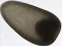
Although this might look like a picture of a flying saucer it is actually a lateral view of a trochophore larva of a polychaete from the genus Polygordius. This larva (and many others like it) were collected from the plankton in Charleston, Oregon on May 10th. The Polygordius trochophore was the first trochophore larva described in the literature (Hatschek 1878), even though it is quite unusual, as far as trochophore larvae go (Rouse 1999). Trochophore larvae, in general, are characterized by having an equatorial ciliary band, called the prototroch. You can clearly see the prototroch in the first picture as a yellow band that divides the larva into two regions - the upper episphere, and the lower hyposphere. The unusual thing about this trochophore, also known as “endolarva” (Woltereck 1904), is that the segmented juvenile body develops tucked inside the spherical larval body. The larva pictured here has a well developed juvenile body inside the transparent hyposphere. The two red spots at the apex of the episphere  are the eyes.  The greenish/yellow ballon inside the larval sphere is the stomach. Polygordius larvae have a through gut and feed in the plankton. 

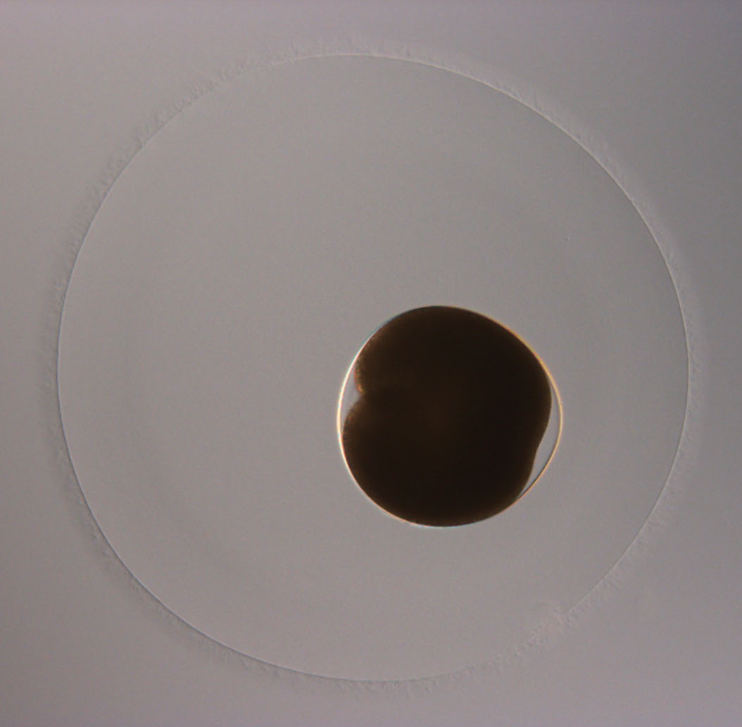
This picture offers a view of the larval apical organ located at the apex of the episphere. The two eyes which are part of it appear black in transmitted light. The apical organ also contains two clear vesicles, which look like statocysts. Statocysts are balance organs found in some larval and adult marine invertebrates. 

Polygordius trochophore is one of the few polychaete larvae that have a catastrophic metamorphosis. I was fortunate enough to witness this process in the lab, and took a picture, as the larva was undergoing metamorphosis (left).  The orange-yellow band at the anterior end (up) is the prototroch. The process of metamorphosis begins with the juvenile suddenly extending out of the hyposphere. The larval body (including the prototroch) is then resorbed.  In this larva the process was more gradual than in some others of this genus - the episphere was resorbed gradually over the course of a few days, and even after a week one could still detect the remnants of the prototroch, even as the now juvenile worm crawled around the bottom of the culture bowl. 

Hatschek B. 1878. Studien über Entwicklungsgeschichte der Anneliden. Ein Beitrag zur Morphologie der Bilaterien. Arbeiten aus dem Zoologischen Institute der Universität Wien und der Zoologischen Station in Triest. 1: 277–404. 

Rouse G. 1999. Trochophore concepts: ciliary bands and the evolution of larvae in spiralian Metazoa. Biological Journal of the Linnean Society. 66: 411–464. 

Woltereck R. 1904. Beiträge zur praktischen Analyse der Polygordius-Entwicklung nach dem “Nordsee”-und dem “Mittelmeertypus”-Typus. I. Die für beide Typen gleichverlaufende Entiwicklungsabschnitt: Vorm E ibis zum jungsten Trochophore-Stadium. Arch. Entw.Mech. Org. 18: 377-403.

Monday, May 28, 2012

Holothuroid doliolaria

I found this gastrula in a plankton sample taken from the Charleston marina docks on May 17th.  We speculated that it may be a sea cucumber (class Holothuroidea; phylum Echinodermata) because of its unusual shape.  One can see in this picture that the gastrula is longer than wide and tapers toward the animal pole (upper left).  In holothuroids, gastrulation begins by invagination whereby cells at the vegetal pole (bottom right) fold into the blastocoel (the cavity within the embryo) as a layer, rather than ingress individually (McEuen 1987).  One can see these invaginating cells in this gastrula as an opaque area.  This embryo later confirmed our suspicions and developed into a lecithotrophic (non-feeding) doliolaria (planktonic larva of some holothuroids). 

Doliolariae are shaped like a barrel (hence the name) and are ciliated. Cilia may cover the entire surface (uniform ciliation) or form 2-5 discrete transverse bands.   Ciliated bands often develop from an initially uniformly ciliated area of epidermis (Miller 2001).  On May  23rd (left) two ciliary bands were present in this larva in addition to a uniformly ciliated anterior area (left on this image). 

By May 25th, the uniformly ciliated region had developed into yet another discrete band of cilia (three in total), seen in the third photograph.The duration of the doliolaria larval stage is species specific, ranging from six to thirteen days in the local species with described development (Miller 2001). The end of planktonic period is marked by the protrusion of five primary tentacles from the larval vestibule.  One can see several curved projections in the larval epidermis (upper left in the third image) through which these tentacles will eventually emerge. At that point the larva will be called a pentactula.

Although sea cucumbers are relatively soft bodied, internal calcareous ossicles are used for structure and support.  In this polarized light image, one can see a small ossicle as well as a larger ring of ossicles that will surround the pharynx in the adult sea cucumber (Ruppert et al. 2004). 

Miller, B. 2001. Echinodermata. In: An Identification Guide to the Larval Marine Invertebrates of the Pacific Northwest. Edited by Alan Shanks. OSU Press, Corvallis.

McEuen, SF.  1987.  Echinodermata: Holothuroidea.  In: Reproductive and Development of Marine Invertebrates of the Northern Pacific Coast by Megumi Strathmann.  University of Washington Press, Seattle. 

Ruppert, E. E., R. S. Fox and R. D. Barnes.  2004.  Invertebrate Zoology: A Functional Evolutionary Approach.  Brooks Cole, Belmont, CA.

Identifying crustacean nauplii

The first picture is that of a copepod nauplius from a plankton tow collected at the mouth of Coos Bay, OR on Saturday, May 5th. The nauplius is the first larval stage of many crustaceans (although many species pass through this stage while still enclosed in the egg capsule). Nauplii are characterized by having three pairs of appendages: a pair of uniramous (not forked) antennules, and a pair of antennae and manidibles, both of which are biramous (i.e. forked). Nauplii are some of the most common organisms one encounters while sorting plankton samples. In Coos Bay we often encounter the nauplii of both barnacles and copepods.

In the second picture we see the nauplius of a barnacle.  Typically barnacles go through four to six naupliar stages depending on species. The distinguishing characteristic of a barnacle nauplius is the pair of fronto-lateral horns at the anterior end. Copepod nauplii do not have any such horns. Being able to distinguish between the two kinds of nauplii is important because both have a biphasic life cycle, but copepods are holopelagic (meaning they spend the entire life cycle in the water column) whereas barnacles are benthopelagic (adults are benthic, while larvae are planktonic) so different ecological questions can be addressed by studying these two kinds of organisms.

After the nauplius barnacles go through an additional larval stage called the cyprid (left). The final nauplius stage molts into the cyrpid stage, which is non-feeding. Cyprid larva has a pair of large appendages at the anterior end (left side of this photo) called antennae  - one can see these sticking out from the carapace. The cyprid will use these to “walk” around on the substrate while  looking for an ideal spot to settle. Once it finds a spot it will cement itself to the substrate and undergo metamorphosis into an adult (permanently attached) stage.

Saturday, May 26, 2012

Brachiolaria larva of Pisaster ochraceus

Pictured here is a dark field image of a four-week old Pisaster ochraceus (a kind of sea star) brachiolaria larva that is 2.35 mm long. This is a ventral view with the anterior end up. The central V-shaped structure is the mouth, which leads into esophagus (a clear tube just below), which in turn leads to the stomach (light pink oval shape below), and finally the intestine (dark pink tube), which opens outside via an anus (out of focus) on the ventral side. The convoluted circumoral ciliary band is particularly obvious (clearly in focus) along the sides of the larval body in the middle part of the image. It is divided into the pre-oral (anterior to the mouth) and the post-oral (posterior to the mouth) portions.  

The second image is a rare side view (left side) of the larva. Located near the anterior portion of the larva is the attachment complex that characterizes the difference between the brachiolaria and the preceding bipinnaria larval stage. Below is a close up of this attachment complex. There are three brachiolar arms capped by the dark colored suckers that test the substratum and provide temporary adhesion during settlement. These arms are differentiated from other larval arms by containing an extension of the anterior larval coelom (which
one can clearly see in the second picture). The dark circle between the two bottom arms is the adhesive disk which contains glands that secrete a cement-like substance for more permanent attachment during metamorphosis.

The last image is a close up of a developing spicule located just near the larval stomach. See an earlier post by Lara Macheriotou for context. This is interesting because asteroids lack the ability to make larval spicules, unlike the pluteus larvae of echinoids and ophiuroids that have a calcareous skeleton.  See a post by Nick Hayman about the underlying developmental mechanism. The spicule pictured here is part of the developing juvenile rudiment, and will be incorporated into the adult skeleton.

Sipunculid pelagosphera larva

This sipunculid (peanut worm) pelagosphera larva was found in an open ocean plankton sample that was collected during our class boat trip on April 17, 2012 – a few miles south of the mouth of Coos Bay, OR. Pelagosphera larva is found only in the phylum Sipunculida. They develop from trochophore larvae and can be lecithotrophoic or planktotrophic. I have kept this larva for a few weeks and fed it unicellular algae Dunaliella tertiolecta and Rhodomonas lens.  Since there is only one known species of peanut worm that has a long-lived, planktotrophic pelagosphera larva in the Pacific Northwest, this larva likely belongs to Phascolosoma agassizii.

The body of this pelagosphera larva can be divided into four regions: the head, the mid-region that includes the metatrochal ciliary band, the trunk, and the terminal organ.  The head, mid-region and terminal organ can be simultaneously retracted into the trunk (left) when the larva is disturbed. A cuticle covers the epidermis of the entire larval trunk. This pelagosphera has numerous cuticular papillae covering the entire trunk. The digestive system can be seen through the cuticle and is indicated by the green hue in the esophagus, stomach and intestine. 

The second picture (left) shows the pelagosphera with extended head, and the metatroch, which these larvae use to swim. The third picture shows the larvae with the head and lip extended. The head is covered by cilia on the ventral surface and divided into two ventral lobes by a ciliated groove that leads into the mouth. The lip is just below the head and is typically covered with cilia on the medial and proximal surfaces. The lip along with lip glands, gland cells of the oral cilature and buccal organ are believed to be involved in feeding.

The terminal organ (not shown) is usually retracted when the larva is swimming or inactive. The primary function of the terminal organ seems to be attachment to the substratum. I was not able to take a picture with the terminal organ of the pelagosphera visible.

Jaeckle, W & Rice, M. 2002. Phylum Sipuncula. In: Atlas of Marine Invertebrate Larvae. Edited by Craig M Young. Academic Press.

Johnson, K. 2001. Sipuncula: The Peanut Worms. In: An Identification Guide to the Larval Marine Invertebrates of the Pacific Northwest. Edited by Alan Shanks. OSU Press, Corvallis.

Thursday, May 24, 2012

Unknown Advanced Actinotroch

This picture shows a lateral view of an unknown actinotroch larva of a phoronid worm that was found on May 10th in plankton sample taken off the F Dock in the Large Boat Basin of Charleston, OR.   This picture clearly shows a ring of tentacles (called the lophophore) at the anterior end of the larva (up). The lophophore is used for feeding and each tentacle is ciliated. The oral hood is visible just anterior to the lophophore. It is pulled over the mouth, much like a hood, and is used in feeding as well. This picture also shows the telotroch,  a ciliated ring at the posterior end of the larva which is used for swimming.

The second picture is a ventral view of the same larva. The telotroch, lophophore, and oral hood are clearly visible. It also shows the achtinotroch’s digestive tract. The stomach is the large internal cavity, and the narrow, tapered end toward the postrior is the intestine. The structure that is wrapped around the posterior part of the stomach (partially outlined by yellow pigment granules) is the metasomal sac. Metasomal sac grows inside the larva, is everted at metamorphosis and becomes the trunk of the juvenile. For a view of an early metasomal sac, see an earlier post by Mark Inc.

The third photo is a dorsal view of the larva. The metasomal sac is now on the far side of the stomach, though it is still visible. The anterior-most end of the sac opens to the outside via the metasomal pore. The sac is everted through this pore. The mouth of the larva is visible as a triangular shape at the anterior end, just below a cluster of pigment granules. It is inside the oral vestibule formed by the oral hood.

Nephtys sp. metatrochophore

This is a metatrochophore larva of a nephtyid polychaete (family Nephtyidae).  In April and May 2012, we frequently encountered these larvae in plankton samples from Coos Bay, OR.  These larvae are propelled by two ciliary bands - an anterior prototroch and a posterior telotroch. The prominent red pigment bands near the prototroch and pygidium (the posterior-most segment) inspired me to look at them more closely.  Upon closer inspection, one can see striking blue (!) pigment in the lining of the gut.  Also in the gut, is this larva’s most recent meal, a centric diatom.  We determined that this larva belongs to the genus Nephtys due to the distinct red and blue pigments mentioned above as well as the dome shaped episphere and a single pair of red eyes (Crumrine 2001).  

In one week, this planktonic larva turned into a benthic juvenile polychaete (left) displaying additional characteristics of Nephtys species.  These predatory worms commonly found burrowing in mudflats use an eversible pharynx armed with a pair of jaws (visible in the third picture as two glowing arrowheads) to capture and subdue prey such as mollusks, crustaceans or other polychaetes.  Furthermore, we noticed that the juvenile possessed an anal cirrus (looks like a small sphere on the pygidium in the bottom photograph).  The presence of an unpaired anal cirrus is  characteristic of Nephtys species, although it is often lost when adults are handled.  

There are more than six species within this genus in the NE Pacific, but N. caeca and N. caecoides are most common (Rudy and Rudy 1983).  To identify species within this genus, one must examine the interramal cirri (slender projections between parapodial branches) on the adult parapodia (Carlton 2007), which was not possible for this juvenile stage.    

Crumrine, L.  2001.  Polychaeta. In: An Identification Guide to the Larval Marine Invertebrates of the Pacific Northwest.  Edited by Alan Shanks.  OSU Press, Corvallis.

Carlton, J T.  2007.  The Light and Smith Manual:  Intertidal Invertebrates from Central California to Oregon 4th Edition.  University of California Press, Berkeley. 

Rudy, P and L Rudy.  1983.  Oregon Estuarine Invertebrates: An Illustrated Guide to the Common and Important Invertebrate Animals.  Oregon Institute of Marine Biology, Charleston, OR.   

Metamorphosed juvenile of Strongylocentrotus purpuratus

These pictures show a newly metamorphosed juvenile of the purple sea urchin Strongylocentrotus  purpuratus, a common intertidal organism in Oregon.  Take note of the red pigment granules and the green coloration of the internal structure though the transparent skeletal plates.  Purple urchin pluteus larvae swim and feed in the plankton for several weeks prior to settlement on the seabed and metamorphosis into the juvenile stage. This particular specimen was cultured in the lab from gametes collected from induced spawning via injection of adults with a KCl solution and fertilized in vitro on April 3, 2012.  Like the adults, the juveniles of this species are benthic feeders and have acquired traits adapted to their environment, for example, tube feet, and juvenile (splayed) and adult (pointed) spines.  At this early stage, the juvenile possesses four adult spines per interambulacrum, two juvenile spines, a single  unpaired primary podium per ambulacrum, and five to eight additional juvenile spines located on the aboral surface (Miller and Emlet, 1999).  The juvenile spines are four-pronged, shorter, and located more orally compared to the longer, barbed adult spines.

The second image is a close up of the oral surface of the juvenile, showing the five tube feet and the transparent skeletal plates.  After settlement, the juvenile is not able to feed for 5 to 6 days due to an incomplete gut.  

Leahy, PS.  1986.  Laboratory culture of Strongylocentrotus purpuratus adults, embryos, and larvae.  Methods in Cell Biology, 27, 1-12.         

Miller, BA., and Emlet RB.  1999.  Development of newly metamorphosed juvenile sea urchins (Strongylocentrotus franciscanus and S. purpuratus): morphology, the effects of temperature and larval food ration, and a method for determining age.  Journal of Experimental Marine Biology and Ecology. 235(1): 67-90.

Thursday, May 3, 2012

Hoplonemertean embryo and larva

This is a hoplonemertean (ribbon worm) embryo. I collected several of such embryos from plankton obtained off of a dock in Charleston, OR on February 8, 2012. In general, nemertean eggs cleave circumferentially, but in some species with large yolky eggs, the cleavage furrow cuts in from one side. This is referred to as “unilateral cleavage”. The embryo shown here is doing just that - it is in the process of dividing from one to two cells, and the cleavage furrow is deeper on one side than on the other.  Many hoplonemerteans encase their eggs in some sort of envelope, or chorion. The embryo shown here has an inner chorion (which is tight around the embryo) and a larger outer chorion. 

The second picture shows a uniformly ciliated multicellular embryo after one day of development. The embryo rotates inside the egg chorion at this stage. It also has an apical tuft of longer cilia (seen at about 5 o’clock). A clear vesicle inside the chorion at about 2 o’clock is likely a polar body (a product of meiosis). The larvae that hatched from these eggs (after 4 days of development) had a transitory larval epidermis which they shed when slightly compressed under a coverslip. 

The third picture shows two ciliated cells of transitory larval epidermis left behind by a larva that is just about to exit the frame (at upper left). Other hoplonemertean larvae are known to have similar transient epidermal covering; in some it is reabsorbed, in others it is shed (Maslakova & von Döhren 2009; Hiebert et al. 2010).

The bottom photograph is of a week-old larva. It is uniformly ciliated and possesses a prominent apical tuft, a caudal cirrus, three pairs of eyespots, cerebral ganglia (large clear areas), gut, and a proboscis. It is essentially a planktonic juvenile. This type of development is referred to as “direct” because the planktonic stage (if present at all) possesses a body plan essentially similar to that of the adult (unlike the nemertean pilidium larva).

Hiebert LS, Gavelis G, von Dassow G and Maslakova SA. 2010. Five invaginations and shedding of the larval epidermis during development of the hoplonemertean Pantinonemertes californiensis (Nemertea: Hoplonemertea). Journal of Natural History. 44: 2331–2347.

Maslakova SA, von Döhren J. 2009. Larval development with transitory epidermis in Paranemertes peregrina and other hoplonemerteans. Biological Bulletin. 216:273–292.